Digital X-Rays
Clear imaging for precise dental diagnosis.
What Can Dental X-Rays Show?

Dental X-rays are vital in helping the dentist and hygienist see areas above and below the gum line that are not visible with the naked eye, such as:
Traditional dental X-rays use a small piece of film to take a photo of the teeth. The film is then run through a series of chemicals to allow the photo to form.
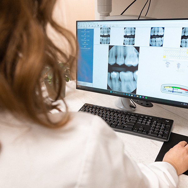
Digital dental X-rays do not use film. They use a sensor in place of film. The sensor is connected to a computer, and the photo is immediately visible on the screen.
What Are the Advantages
of a Digital X-Ray?
There are many benefits to modern radiographs. Here are the top three:
-
Safety
Many offices prefer to use digital X-rays for a number of reasons. Primarily, there is much less radiation needed to take a digital X-ray. Because the sensor is so sensitive, only a minimal amount of radiation is needed to see the picture. Additionally, no dangerous chemicals are needed for digital X-rays, making them more environmentally friendly. Lastly, digital X-rays are stored on a computer; they will not fade or get lost over time and can be easily shared with specialists if necessary.
-
Provides a Unique Record
Every patient’s radiographic needs are different. Most dental offices will not accept a new patient without some form of X-rays present in their record, either from their former dentist or ones that may be taken at their first visit. Once an initial X-ray record is established, records are updated periodically at check-up visits.
-
Prevent Costly Procedures
Patients who are at high risk for cavities, have many restorations present in their mouth or have a history of gum disease may need to have X-rays more frequently. By closely monitoring X-rays, conditions such as cavities may be caught early and allow for less invasive and less costly procedures. Patients who do not have any or have few restorations, good oral hygiene, and no history of gum disease may not need X-rays updated as regularly.

Types of Digital Dental X-Rays
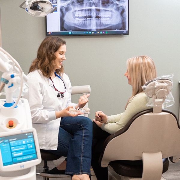
Panoramic X-rays give an overall view of the entire mouth and surrounding anatomic structures in one photo. The dentist may ask for this type of photo to view the position of impacted teeth or cysts, assess the TMJ, or view forming permanent teeth below the primary teeth in children. While these photos are good at giving a general view of the mouth, they do not show small cavities that may be present or forming.
A Full mouth series of X-rays consist of 16 to 18 smaller X-rays that give a closer view of the teeth and bone. A full mouth series is made up of periapicals (which show the entire root and tooth) and bitewings (which show how the teeth fit together in a bite). These smaller photos allow the dentist and hygienist to assess bone levels, check for cavities that may be forming, the integrity of any restorations present in the mouth, and cracks in the teeth or roots.
-
Monday - 9:00am–5:00pm
-
Tuesday - 8:00am–5:00pm
-
Wednesday - 8:00am–5:00pm
-
Thursday - 8:00am–5:00pm
-
Friday - 8:00am–5:00pm


